
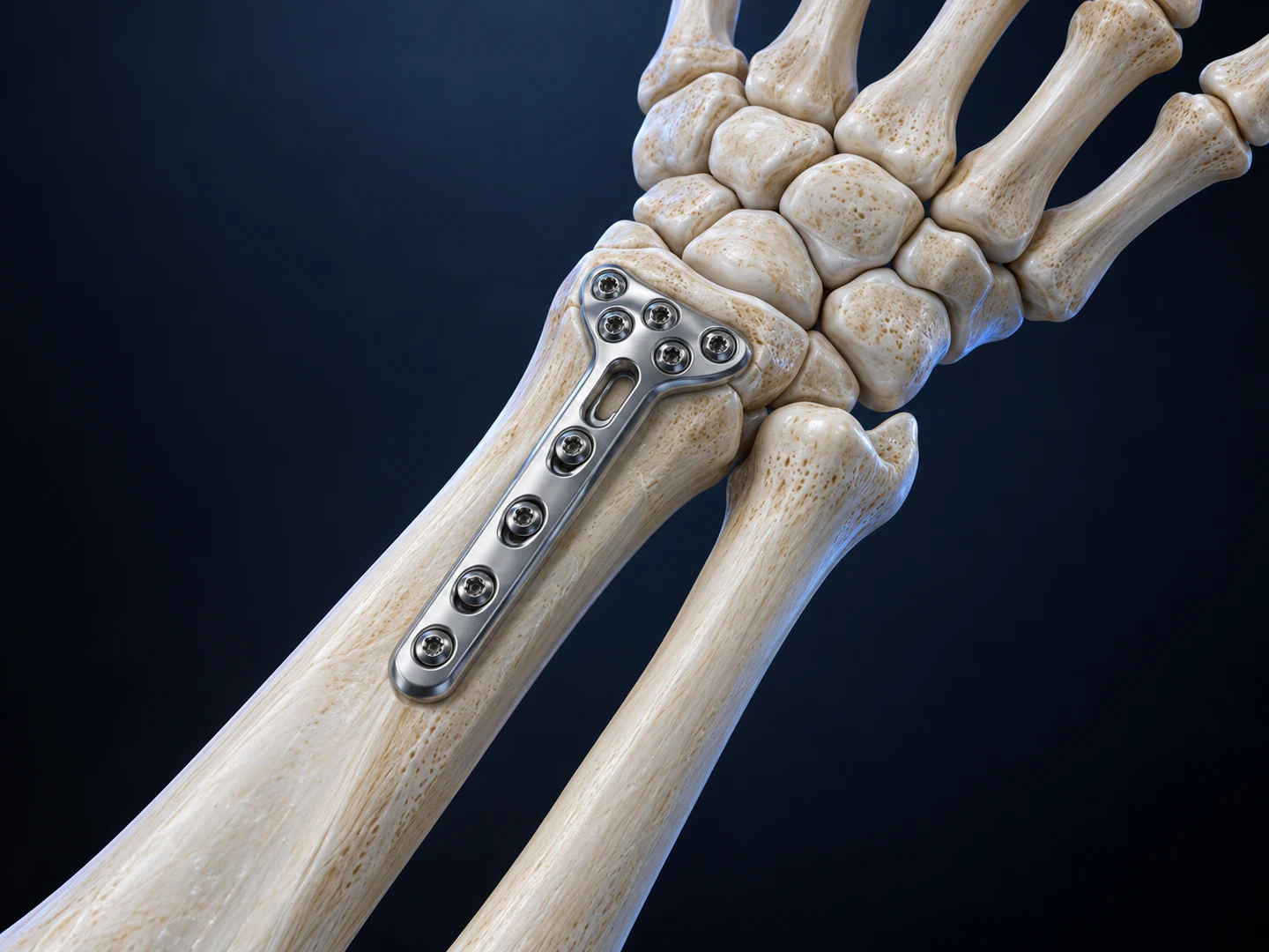
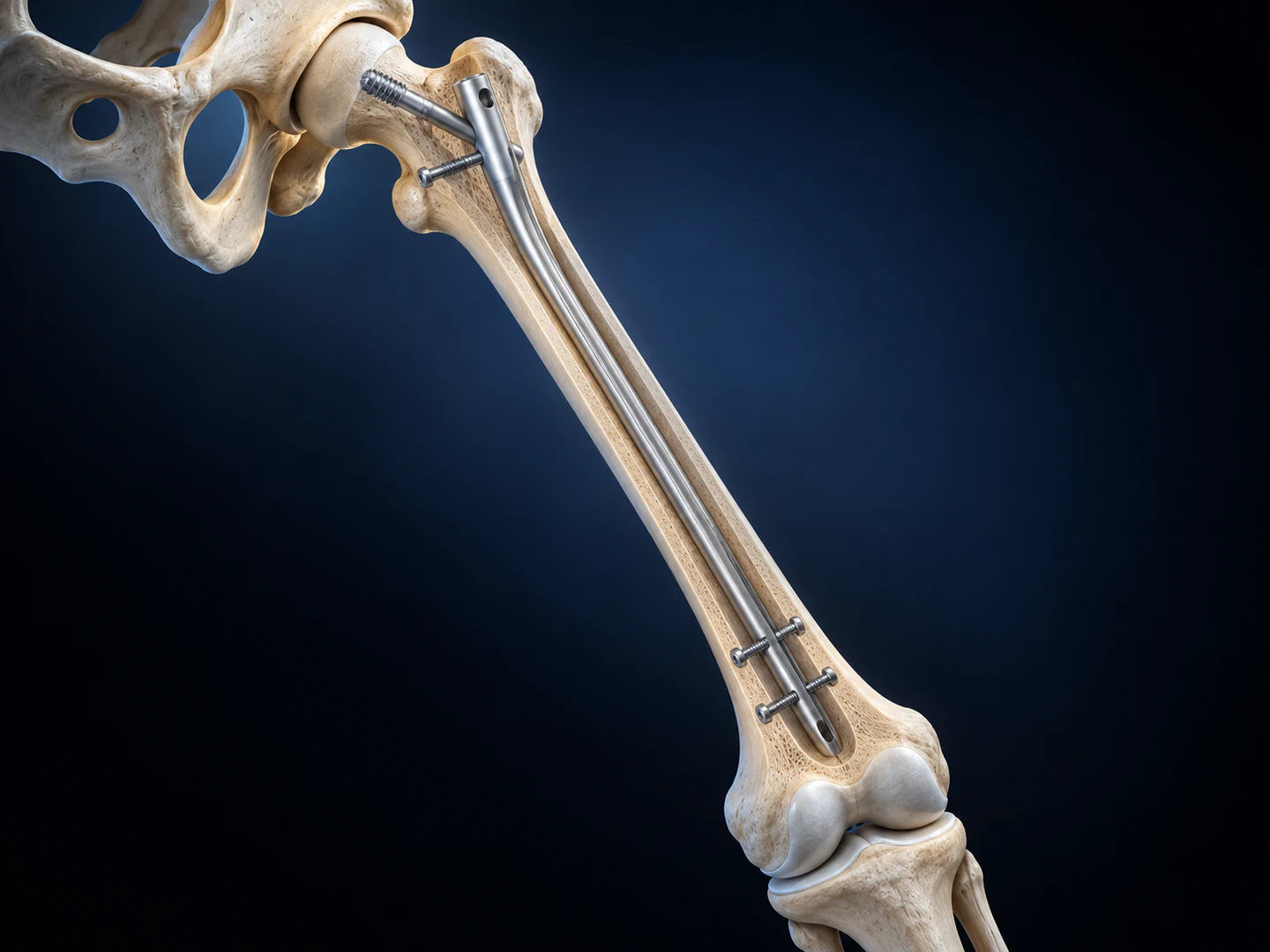
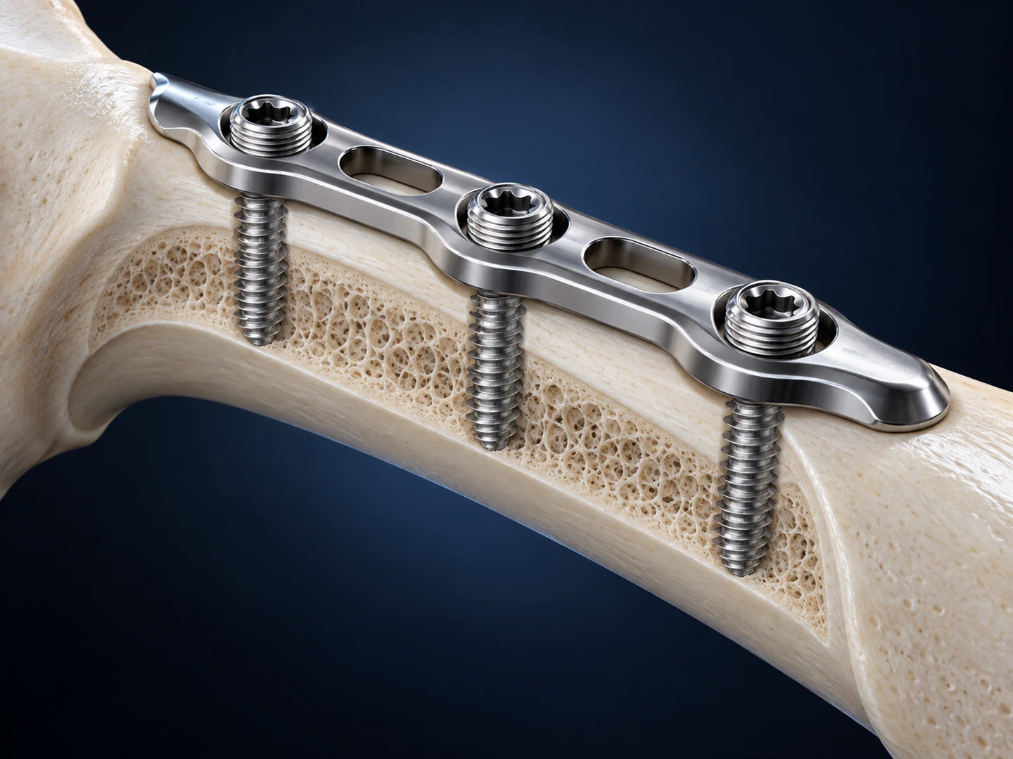
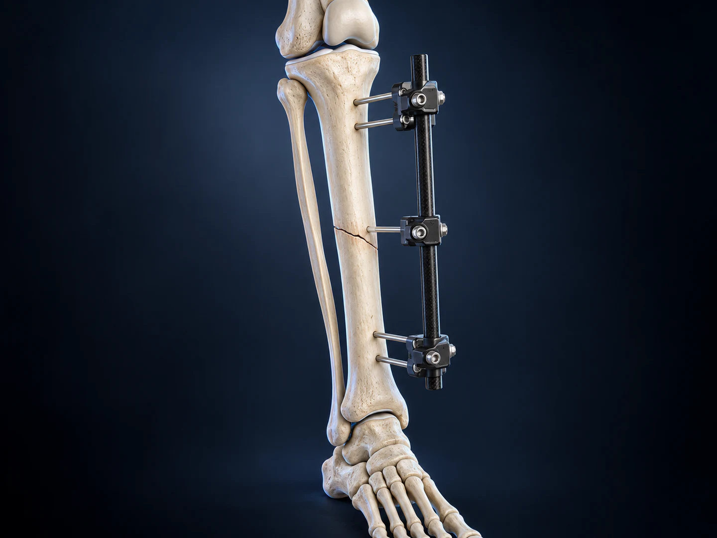
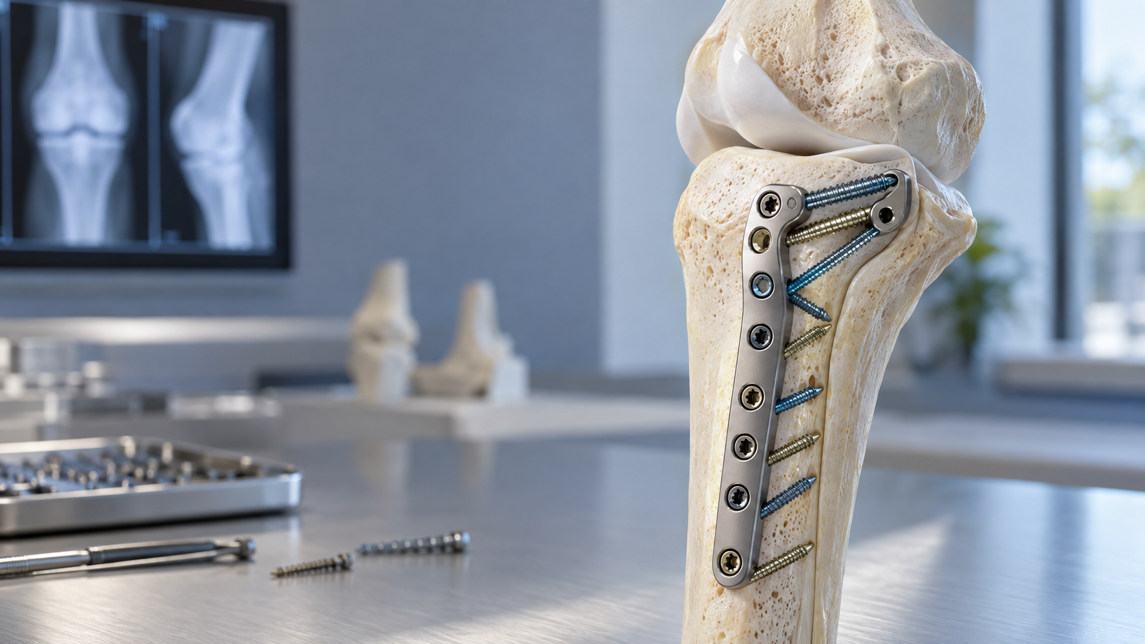
Periarticular fractures rarely present with a single ideal screw path. Fragment size, bone quality and joint geometry often require controlled flexibility while the plate itself remains stable.
Controlled freedom at the plate
Variable-angle locking technology allows the surgeon to direct screws within a defined cone while preserving a locked screw-plate interface. This can help capture individual fragments, avoid the joint and work around existing hardware.
Planning still matters
Angular freedom does not replace reduction quality or sound construct planning. Screw trajectory, length, density and plate position must be considered together, particularly in osteoporotic metaphyseal bone.
Practical workflow
- Confirm plate position before final locking.
- Use provisional fixation to protect reduction.
- Select trajectories that support key articular fragments.
- Verify every screw with appropriate imaging views.
Used deliberately, variable-angle fixation gives the surgical team another way to balance fragment capture with construct stability.
Where trajectory flexibility adds value
In periarticular fixation, the most useful trajectory is not always perpendicular to the plate. A fixed-angle path may intersect a fracture line, miss a small articular fragment or conflict with another screw. Controlled angulation gives the surgeon room to respond to those constraints while staying within the system’s validated locking envelope.
This is especially relevant around the distal radius, proximal tibia and other metaphyseal regions where fragments are small, bone quality may be reduced and the available corridor is limited by the joint surface. The goal is not maximum angulation. The goal is the smallest purposeful adjustment that improves fixation without compromising safe placement.
Construct strategy in weaker bone
Osteoporotic bone changes the way loads move through a construct. More screws do not automatically create better fixation. Plate length, working length, screw distribution and support beneath the articular surface all influence stability. Variable-angle screws may improve fragment capture, but they still depend on appropriate plate position and adequate bone purchase.
A balanced construct commonly combines strategically directed periarticular screws with diaphyseal fixation that respects the intended mechanical environment. Surgeons should follow the approved technique and avoid clustering trajectories in a way that weakens bone bridges or creates avoidable conflicts.
Imaging and verification
Freedom of angulation increases the importance of imaging discipline. Every trajectory should be checked for joint penetration, unwanted convergence and appropriate length. Standard views may need to be supplemented by anatomy-specific projections when the joint surface is difficult to assess.
- Seat the plate and confirm reduction before committing to final trajectories.
- Use guides and calibrated instruments supplied for the locking system.
- Check the drill path before measuring and inserting the screw.
- Review the complete construct, not only the most recently placed screw.
Instrument handling and locking integrity
A variable-angle interface has a defined range. Driving outside that range, cross-threading the head or repeatedly redirecting after engagement can reduce locking performance. Clean recesses, aligned drivers and controlled final tightening help protect the screw head and plate interface.
The operative team should also verify that the correct screw family, drill guide and driver are on the field. Similar-looking components are not necessarily interchangeable across systems.
Clinical takeaway
Variable-angle locking is most valuable when it supports a clear fixation objective: capturing a key fragment, avoiding the joint, working around existing hardware or improving subchondral support. It should be used as part of a complete plan that begins with reduction and plate position and ends with systematic imaging verification.